


 Global Sourcing Spotlight: Golf, Friedman, and the Benefits of Global Sourcing
 Nolan’s Notes: Coming to Terms With AI
 The Knowledge Base: A CM’s Perspective on Box Build Practices
Video Microscopes Meet Inspection Requirements
December 31, 1969 |Estimated reading time: 5 minutes
Acquiring information from video images is a common phenomenon. It is a natural progression to extend video imaging to magnification of small objects, such as electronic components on a printed circuit board (PCB) with video microscopes that display images on monitors or flat panel displays.
By Mark C. Hogrebe
Instead of using only light and optics to enlarge an object, video magnifiers use a small image sensing device in combination with optics, light, electronic circuits and computer software to create magnified images.
Conventional Microscopy
Looking into the eyepieces of a conventional microscope for the first time is not an easy task. Clarity of image and depth perception are achieved by the experienced user, but the beginner may have a difficult time coordinating visual paths.
Although the eyes adapt to looking through eyepieces, it requires continual effort to align and integrate the visual information. Over time, this sustained effort produces fatigue. The negative impact on the person of prolonged microscope work has been widely documented and studied. A high percentage of health issues has a significant negative impact on employee productivity, as well as increasing worker compensation costs.
Video microscopes project the magnified image on a monitor, which means more than one person can view the magnified image. Demonstration of procedures and inspection techniques can be made to groups who view the monitor simultaneously.
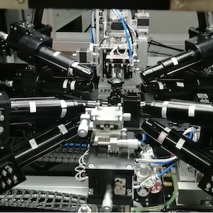
 Figure 1. Camera on boom stand with separate monitor.
Video microscopes use electronically processed images that can be saved as movies or still images and used for documentation in quality control. Typical video microscope configurations consist of a camera connected to a monitor with a cable. The camera is mounted on a "boom" arm/stand (Figure 1) or attached to a conventional microscope (Figure 2).
 Figure 2. Camera on microscope with separate monitor.
Increasing monitor size will make the magnified images look larger, but will not improve image resolution produced by the optics and camera. A video microscope system that uses a large monitor may report high magnification power based on screen size, but may actually have low resolution and poor clarity. The camera and optics determine image quality and resolution at higher magnification levels.
- Integrated for direct line of sight. Integrating the camera and monitor into a single unit helps achieves "direct line of sight" viewing. This is a concept inherent in many glass magnifiers because they maintain the natural line of sight between the eyes and hands. A compact, integrated video microscope approximates the direct line of sight by permitting simultaneous viewing of the original object and magnified image. This relationship enhances perspective, comfort and hand-eye coordination (Figure 3). The greater the distance and lack of alignment between the object and image, the more difficult it is for the user to coordinate their eyes and hands.
- Screen size. A large 15 or 17" monitor may make the image look bigger without an in-crease in resolution and at the cost of occupying valuable workstation space. An ideal size is a 6 to 8" screen that yields high resolution while displaying a visual field that is comfortable to process at a distance of 12 to 18". This distance from eyes to screen is determined by the user's arm reach.
- Working distance. The distance from the bottom of the unit and the work surface needs to be sufficient to perform tasks. Inspection tasks require less working distance than production or repair; however, users generally prefer 8 to 9".
- Image control. Characteris-tics such as brightness, sharpness, white balance and negative image can be varied to produce the best image for a particular inspection task. Video mi-croscopes should have continuous zooming so that images can be made smaller or larger with the touch of a button, instead of having to change lenses.
- Lighting. Good video microscopes use cameras with automatic gain that adjust for different light levels. Together, an automatic white balance and automatic gain provide a great deal of light control. A variety of lighting solutions are available for different inspection tasks and the video microscope should be able to incorporate them.
 Figure 3. User can see both image and object.
A video microscope also should be adaptable to a variety of magnification tasks in the workspace. One mounted on a sturdy, adjustable arm has significant advantages over systems with heavy weighted bases and separate full-size monitors. An ad-justable arm clamped to a table edge leaves a small footprint on the work surface, and uses no surface space when mounted to a wall or directly to a workstation vertical track. When mounted between two adjacent workstations, the reach of the adjustable arm allows the unit to be used in either location.
SMT Applications for Video Microscopes
In contrast to automated optical inspection (AOI) systems, video microscopes are designed for use in place of conventional microscopes. Typical applications are off-line inspection and rework of PCBs. Operators inspect for bridging, proper solder flow, and component placement, orientation and damage. Since the costs of off-line inspection and rework by an operator are greater than ongoing inspection by existing automated systems, it is extremely important to minimize the time that a PCB is at an operator's workstation. The quicker a PCB can be processed, the lower the inspection or rework cost per unit. Opera-tors are able to process more boards over the course of a shift using an ergonomic video microscope because the onset of physical and mental fatigue are delayed. As fatigue is delayed and lessened, accuracy and productivity are maintained.
Since video microscopes generally allow for a greater working distance between the PCB and the lens, it is much easier to maneuver soldering equipment into place and to hold tools naturally while making repairs. A greater working distance and adjustable height gives the operator the ability to manipulate a PCB in order to inspect components and solder flow from various angles.
In addition to savings from increased operator productivity and flexibility, video microscopes reduce costs by shortening training time. More than one person can view an instructor's technique while working under magnification. Everyone observes a complex solder repair in real time, with no need to alternate views or trade positions and have the instructor attempt to repeat the same actions. Second, new operators do not have to "get used to" looking through a microscope. They can immediately start acquiring content and technique without the learning curve associated with aligning the eyes to the visual paths of microscope optics.
The image capture capabilities of video microscopes make documentation and communication of PCB de-fects and repairs a seamless process. With the desired image on the video display, files can be created for immediate transmission to departments within the facility or to suppliers and customers around the world.
Conclusion
Video microscopes alleviate many factors that contribute to operator fatigue, which leads to decreased efficiency and accuracy. Quicker training with use of video microscopes means employees contribute productively in less time. Easy video capture and documentation allows for fast communication and feedback with suppliers, colleagues and customers.
Mark Hogrebe, Ph.D., president, may be contacted at Dazor Manufacturing Corp., 4483 Duncan Ave., St. Louis, MO 63110, (800) 345-9103, Fax: (314) 652-2069; E-mail: mhogrebe@dazor.com; Web site: http://www.speckfinder.com
Share on:


